
Contrast enhanced-ultrasound to identify potentially viable tissue within the penumbra of human spinal cord injury
Principal investigator
 Christoph Hofstetter MD, PhD
UW Medicine Department of Neurological Surgery
Research team
Matt Bruce PhD, UW Applied Physics Laboratory
Zin Khaing PhD, UW Medicine Department of Neurological Surgery
Study description
Critically reduced blood flow to the contused spinal cord is thought to contribute significantly to additional secondary tissue damage and poor functional recovery. This research team aims to delineate mechanisms underlying local hypoperfusion and to develop treatment strategies to restore physiological blood flow to the injured spinal cord.
This lab has recently developed a novel ultrafast ultrahigh-frequency contrast-enhanced ultrasound (CEUS) technique to assess in real-time the structural and functional integrity of intrinsic vasculature, both large and small vessels, within the spinal cord. This technique provides a topographical map of microvascular disruption and perfusion changes at and near the lesion site and thus allows detection of the hypoperfused penumbra adjacent to the injury center in a rodent model of traumatic spinal cord injury. Successful medical and/or surgical restoration of blood flow to this penumbral tissue will likely improve functional outcomes. Currently, the research team is further developing these CEUS capabilities to translate and advance their understanding in an explorative observational clinical trial utilizing an existing FDA approved contrast agent and ultrasound system.
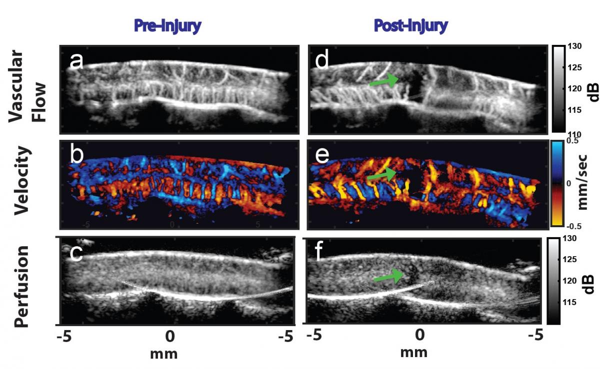 Recently, the research team has enabled contrast-enhanced ultrasound (CEUS) using an FDA approved contrast agents in a rat model of SCI. Their innovative ultrasound imaging is able to detect changes in vascular flow, velocity and even tissue perfusion within the spinal cord. In addition, they are able to detect local changes to the blood flow and vessel morphology after a contusion type spinal cord injury. Importantly, these ultrasound images are acquired very quickly, giving the research team a unique opportunity to examine blood flow changes in real-time.
In collaboration with Philips, a leading provider of ultrasound systems and technology, the research team has contributed to and tested the development of an intra-operative probe in combination with an FDA approved system supporting CEUS in their rat model. The image below illustrates a perfusion deficit in contused rat spinal cord. Following UW Human Subjects IRB approval, they have performed CEUS in several spinal cord surgeries working out the details of optimal contrast agent injection approaches, contrast agent types, and imaging strategies. Below are ultrasound images from a patient with C5 ASIA a traumatic spinal cord injury after a motor vehicle accident. Sagittal T-2 weighted MRI shown multilevel severe impingement of the spinal cord (A). B-mode sagittal ultrasound image (upper panel, B) and contrast-enhanced ultrasound imaging (lower panel B) reveals an area of hypoperfusion which correlates to the neurological level of the patient’s injury.
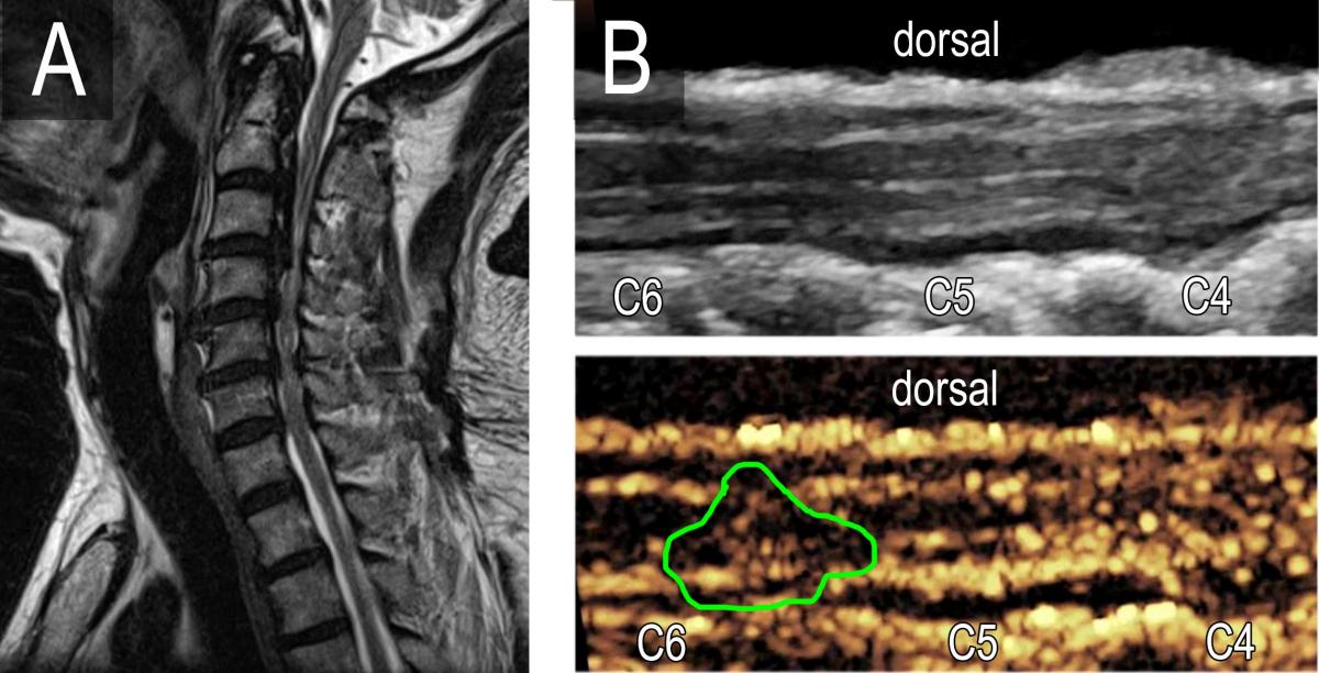 CEU imaging in patients with spinal cord injury. A 68 yo male with C5 ASIA A traumatic spinal cord injury after a motor vehicle accident. Sagittal T-2 weighted MRI shown multilevel severe impingement of the spinal cord (A). B-mode sagittal ultrasound image (upper panel, B) and contrast-enhanced ultrasound imaging (lower panel B) reveals an area of hypoperfusion which correlates to the neurological level of the patient’s injury.